Questions:
1. Why is the small intestine on top of the large intestine? We thought that the large would be below the small.
2. Why is the gallbladder located inside of the liver, and not next to it? We were expecting for the gallbladder to be next to the liver, not in between its lobes.
Interesting Fact:
1. Each section of the small intestine is not independent of the other sections of the small intestine. The small intestine is stored as a single unit, attached by a connecting tissue.
Reactions:
1. Beril was intrigued about how the organs all fit together perfectly inside of the cat's body cavity.
2. Deborah was excited about cutting out the small intestines because the class had learned earlier in the year that they are longer than we would think they are. She was interested in discovering their length.
Difficulties:
1. Deborah had a difficult time cutting out the liver. She and Beril were unsure at first if it was attached to anything else inside of the cat, so it took some time for them to figure out where and how to cut the liver out.
2. Beril and Deborah were not positive about where the Ileocecal valve was located.
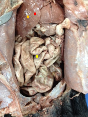 |
| Our view of the liver and small intestines before removing them. |

 |
| We discovered that the Gallbladder was hidden within the liver. |
Red- liver
No comments:
Post a Comment