Tuesday, April 1, 2014

Cat vs. Human Comparison

How are the human and cat digestive systems similar and different?
Similarities
Both have a tongue, salivary glands, pharynx, esophagus, stomach, gallbladder, liver, small intestine, large intestine, rectum, and anus.
Both can digest meat.
Both have molars and premolars.
Both secrete saliva in order to moisten, soften, and chemically break down starches in food.

Differences
Humans
Cats
Omnivores
Obligate carnivores-Can only get nutrients from meat and bones. Can tolerate only small amounts of vegetation & plant matter (only to aid in digestion & to make hairballs)
Humans obtain necessary nutrients and vitamins from plants and animals.
Plants are unnecessary for a cats’ diet. A healthy diet for a cat involves high levels of protein and low levels of carbohydrates.
Humans need carbohydrates to make energy.
Cats need no carbohydrates for energy; it is even unhealthy for them to have carbohydrates such as wheat, corn, and soybeans because they make digestion more difficult.
Slower metabolism than cats and meat must be cooked to eliminate harmful bacteria. Slow metabolism= a higher chance for harmful bacteria to grow (for example: salmonella)
Higher metabolism than humans. Also, a higher metabolism decreases the risk of illnesses from harmful bacteria.
Digestive juices in a human are less acidic than a cat because we do not need to break down bones because we do not eat them.
Digestive juices are highly acidic. This is to help it break down the bones of its prey that it may have eaten.
A human’s liver has 4 lobes
A cat’s liver has 6 lobes
A human large intestine includes the ascending, transverse, and descending colon.
A cat’s large intestine does not include an ascending colon.
Humans do not require the same levels of taurine to function and maintain proper health like cats do.
Cats require high amounts of taurine (an amino acid) in order to maintain proper vision, digestion, heart functions, & immune system.
Tongue is used only to mechanically break down food.
Tongue is used to not only break food down mechanically, but for self-grooming purposes with the papillae.
Teeth are blunter than cats, and are used mainly to just help tear up meat.
Cat teeth are sharper than human teeth, and are used to hold and kill prey.
Premolars and molars are used primarily for grinding up food.
Premolars and molars are used primarily cutting up and tearing food.
Incisors are used to cut up food.
Cat incisors are smaller than a human’s and are used for grooming.
Humans have a wider range of movement for the jaw than cats do, including being able to move the jaw both side to side and up and down.
Cats can only move their jaw vertically, not side to side.
Adult human has 32 teeth.
Adult cat has 30 teeth.
Here is a better view of how the digestive system of the two compare and contrast:

Cat teeth
Human teeth

The Human Digestive System

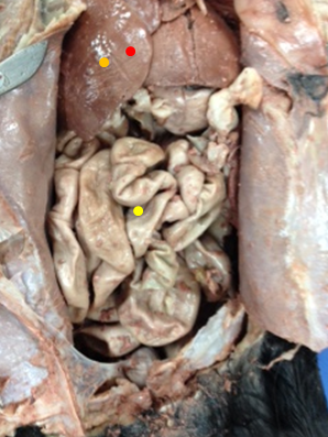
A clearer look at the internal organs...

Monday, March 31, 2014

The Digestive System


  • Mouth
Functions:
    -mastication
    -deglutition
    -start of digestion (starch)
    -self-cleaning
    -holding food
Secretions:
    -Saliva from salivary glands (parotid, sublingual,  
     submandibular)- prepare food for swallowing, prevent mouth 
     from drying, and start the breakdown of starch
    -Amylase- digests starch
Histology: stratified squamous epithelium 
  • Stomach
Functions:
     -temporary food storage
     -mechanical breakdown of food
     -breakdown of chemical bonds by acids and enzymes
     -production of intrinsic factor needed for vitamin B12
      absorption
     -begins the breakdown of proteins 
Secretions:
      -mucous- protects the stomach lining
      -gastric juices- softens meat and bones, kills bacteria in food,
       activates protein-digesting pepsinogen 
      -Hydrochloric acid
Histology: simple columnar epithelium with mucous cells
  • Small Intestine
Functions: 
      -completes the digestion of food
      -absorbs end products into blood and lymph
      -secretes hormones that help control production of pancreatic 
       juice, intestinal juice, and bile
      -controls the amount of fluid and electrolytes lost from the 
       body 
Secretions:
      -paptidase- digest proteins
      -maltase, sucrase, lactase- digest disaccharides
      -lipase- digest lipids 
      -bicarbonate ions- act as emulsifying agents
      -secretin- stimulates the pancreas to secret a fluid high in 
       bicarbonate ions that help neutralize chyme
      -CCK- stimulates the release of bile from the gallbladder and 
       the secretion of digestion enzymes from the pancreas
Histology: simple columnar epithelium 
  • Liver
Functions:
      -produces bile and plasma proteins
      -detoxifies blood
Secretions:
      -bile- lowers the surface tension of fats, forming small droplets
Histology: dense connective tissue
  • Gallbladder
Functions: store and secrete bile into the small intestine in the  
       presence of fatty acids
Secretions: bile
Histology: simple columnar epithelium 
  • Pancreas
Functions: 
      -secretes enzymes into the cavities and blood
      -neutralizes chyme
Secretions:
      -amylase- digest starch into maltose
      -trypsin- digest proteins
      -lipase- digest lipids
      -nucleases- digest nucleic acids
      -sodium bicarbonate ions- neutralize chyme
Histology: simple cuboidal epithelium
  • Large Intestine
Functions: 
      -reabsorb water
      -store wastes
Secretions: none
Histology: simple columnar epithelium 
  • Rectum
Function: temporarily stores feces
Secretions: none
Histology: smooth muscle
  • Anus
Functions: helps feces to exit the body
Secretions: none
Histology: simple columnar epithelium

Day 5 Journal-Time to Brainstorm

On Thursday March 27th, Beril and Deborah thought of several possibilities for their project. At first we were planning on making a journal, but then decided to make a blog. A blog, we concluded, was the best option because we could both work on it from our own homes and because we wouldn't have to print out our pictures and lose a lot of ink. We discussed how we would organize the information together with the pictures and assigned different roles for each other.

On Friday March 28th, Deborah brought her laptop to class so that she and Beril could work on the blog. We opened a Google account and started a blog. We chose the most convenient template and appropriate colors. When we finished with all of the little details, we both started working on writing about day one of the lab.

Saturday, March 29, 2014

Day 4 Journal- That's a Wrap!

On Wednesday March 26th, during period two, Beril held the legs wide open while Deborah continued removing the digestive organs from the body of the cat. After finishing with the small intestines, Deborah cut the stomach out and, together with Beril, examined the inside. Deborah then cut out the large intestines and finally finished the dissection process at the rectum. 

Questions:
1. What volume of food can a cat's stomach hold when at its maximum capacity? 

2. While examining the inside of the stomach, we saw a brown-beige compound in the cat's stomach; was this chyme or digestive juices?

Interesting Facts:
1. After learning about the rugae in the stomach, it was interesting to see how they actually looked and the way the folds were set up in reality.

Reactions:
1. We were uncomfortable putting our hands inside of the cat to remove the deeper internal organs (such as the stomach).

2. We found it strange that the small intestines were made up of a thin layer of smooth muscle. We were expecting for the small intestine to be thicker.

Difficulties:
1. Beril found it difficult to cut the stomach to view the inside because it was very slippery and rubbery.


A closer view of the internal organs
White- diaphragm
Red- stomach
Yellow- small intestine


Stomach when removed from the body.
View of the inside of the stomach and its rugae.
Examining the small intestine.
The rectum (labeled in red): second to last stage of the
digestive system
Lastly: the end of the dissection after removing the internal organs

Day 3 Journal- Surgeons at Work

On Wednesday March 26th, during period one, we removed the cat and its accompanied juices from the bag yet again. Just like the day before, Beril held the legs wide open, while Deborah finished cutting open the abdominal and thoracic cavities. Deborah cut out the liver and Beril helped her to locate the gallbladder that was attached on the inside of the liver. Beril switched off from holding the legs of the cat open, answering questions in the light blue packet, and taking pictures. Deborah then cut out the small intestines to examine and measure its diameter. 

Questions:
1. Why is the small intestine on top of the large intestine? We thought that the large would be below the small. 

2. Why is the gallbladder located inside of the liver, and not next to it? We were expecting for the gallbladder to be next to the liver, not in between its lobes.  

Interesting Fact:
1. Each section of the small intestine is not independent of the other sections of the small intestine. The small intestine is stored as a single unit, attached by a connecting tissue. 

Reactions:
1. Beril was intrigued about how the organs all fit together perfectly inside of the cat's body cavity.

2. Deborah was excited about cutting out the small intestines because the class had learned earlier in the year that they are longer than we would think they are. She was interested in discovering their length. 

Difficulties:
1. Deborah had a difficult time cutting out the liver. She and Beril were unsure at first if it was attached to anything else inside of the cat, so it took some time for them to figure out where and how to cut the liver out.

2. Beril and Deborah were not positive about where the Ileocecal valve was located. 

Our view of the liver and small intestines before
removing them.

We discovered that the Gallbladder was hidden within the liver.

Yellow- gallbladder
Red- liver

Friday, March 28, 2014

Day 2 Journal- Let's See What's Inside

On Tuesday March 25th, we removed the cat and its accompanied juices from the bag. Beril held the legs wide open, while Deborah cu the abdominal and thoracic cavities open. At this stage, we were able to see half of the digestive system, the liver and small intestines, and some of the accessory organs. We then discovered that the stomach and large intestines are hidden behind the liver and small intestines.

Questions:
1. Why didn't we see any bones?

Interesting Facts:
1.The small intestines is on top of, rather than above, the large intestines. 

2.The gallbladder is located within the liver, not next to it. 

Reactions:
1. Beril was surprised about how many layers we had to cut through in order to view all of the internal organs.

2. Deborah's hand began to feel sore from cutting the car because it was difficult at first to remove all of the layers from on top of the organs. 

Difficulties:
1. It was difficult to cut the cat without having one of us holding the legs open, because the cat would not lie flat on its back by itself. 

Our view of the inside of the thoracic and abdominal
cavities after cutting them open.
Red- stomach
Orange-liver
Yellow- gallbladder (in the middle of the liver)
Green- small intestine
Blue- large Intestine (below small intestine)
Purple- rectum (below intestines)

Day 1 Journal- Oh, a Cat!

On Monday March 24th, we worked together to get the cat out of its bag and drain all of the juices in the bag. We needed Woj's help, but were able to clean up the spill. We then examined the cat's exterior to answer the lab questions in the light blue packet, and worked together to carefully put the cat back into the bag.

Questions:

1. What was in the preservatives?

2. There was a small cut in the cats's left leg. We noticed that all three layers of skin were removed from that section of the leg, and that the muscles and veins were showing. We noticed that one vein was a dark shade of blue. Why was the leg cut? Why was the vein blue?


Interesting facts:
1. We found it interesting that cats whiskers help cats with navigation and sensing their environment.


2. The cat's tongue contains papillae for cleaning and eating purposes.


Reactions:
1. Our initial reactions to the wet, smelly, heavy cat was disgust.

2. After taking the cat out of the bag, draining the preservative juices, and examining its exterior, we were more comfortable with dissecting it.

Difficulties:
1. We had a difficult time containing the liquid inside of the bag. As we were removing the cat, the liquid spilled over onto the table and floor. 

The cat when we first took it out of its bag. 
Examining the cat's tongue, the first part of the
Digestive system.